Geologists like me teach students and conduct research with microscopes called "polarizing" or "pol" for short. After development in the late 19th century, pol scopes became critical tools for studying the minerals and structures of rocks (our Earth), which can tell us much about how they formed. Such users are petrologists, who are doing petrography. Polarizing microscopes are also used by mineralogists, geophysicists, soil scientists, crystal chemists, materials engineers, medical researchers, asbestos consultants, and many others including biologists.
The method that can be employed with these instruments is called PLM (polarized light microscopy). "Simple polarizing" scopes are usually just biological microscopes with pol filters, one inserted under the head and another in a substage filter holder. About any microscope can be so modified, including stereo microscopes, but the common type is a compound microscope. Those designed for geological work are also known as "petrographic" (="rock imaging") microscopes, which have accessories to assist mineral studies in thin sections of rocks.
Many other crystalline materials (organic as well as inorganic) produce interesting optical effects with both polarizers in place, and some biological samples can show useful changes of contrast against a dark background.
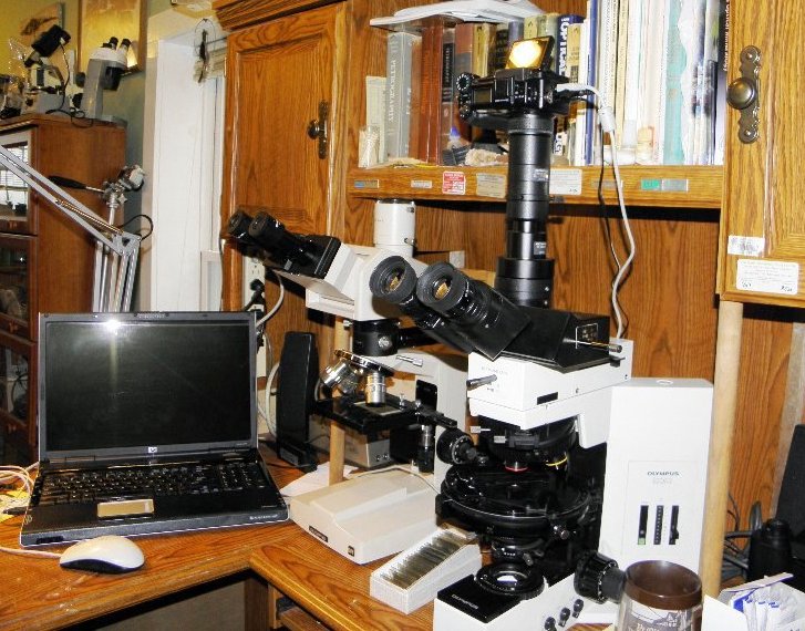
In my research lab, an Olympus BH2 BHSP petrographic polarizing microscope (below) is serving me well, and I am convinced that it is the best of the 20th century "benchtop" pol microscopes. I will use it to illustrate pol scopes.
You might disagree with this assessment, but in my research and business I have used more than two dozen different petrographic microscopes of many brands and models. Actually, I liked them all, but some were better than others in design, function, and quality. Six of the best pol scopes in my experience are the subject of my article in the internet journal called Micscape, also linked below. I have also described some interesting student petrographic microscopes in that journal. Eventually I added a super-wide head and eyepieces with a view field number of 26.5. It includes about 2.65 mm of slide in the view with the 10x objective, or 6.625 mm with the 4x, which is huge. Very nice for scanning coarse grained rock slides, but I had to renovate stuck diopters on these special oculars.
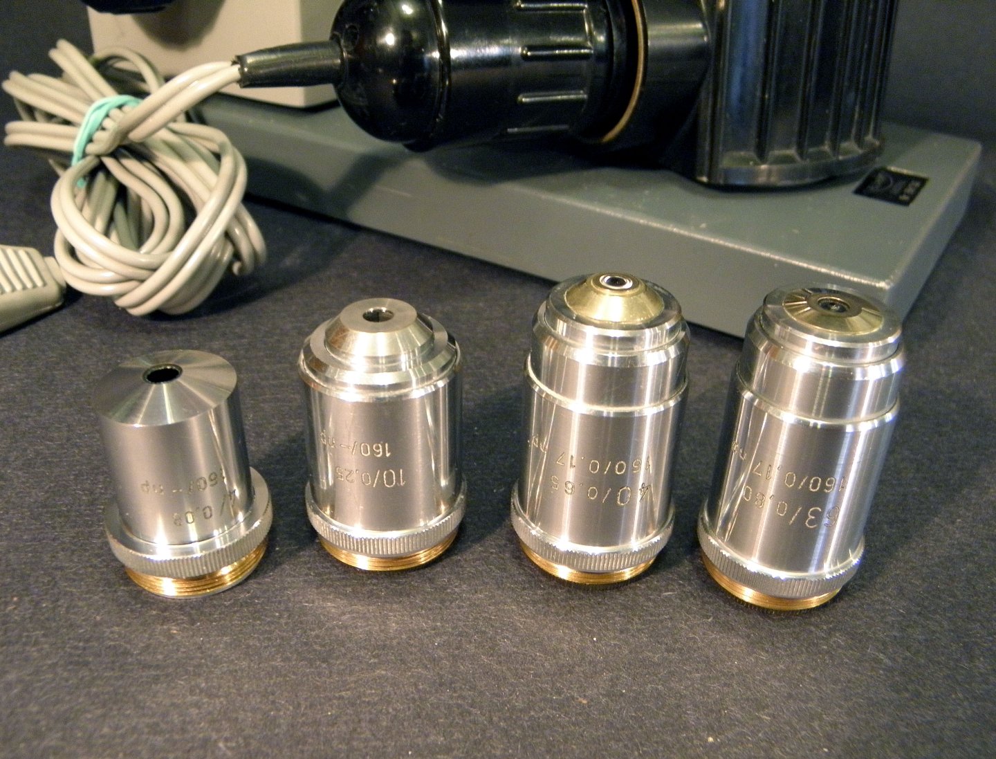
I have also fixed several Zeiss Pol objectives with frozen focusing rings by completely disassembling, cleaning, re-lubricating, and reassembling them. It may not be for the faint-hearted but it is not rocket science either. You do need proper tools.
Another Micscape article describes the path to obtaining my BHSP, including earlier petrographic microscopes that I used for geological research. New 21st century models such as the Zeiss Axio Scope Pol or Olympus BX53P are no doubt even better microscopes than my late 1980s machine, but my budget will never allow such new and very expensive microscopes. In fact, there are a good number of late 20th century microscopes that can meet professional requirements for a modest cost, and also be satisfying to own and use. My personal research microscope is now an Olympus BX50P from the mid-1990s, which has matchless optics and quality about as high as the BHSP -- excellent!
How is a pol scope different?
There is nothing especially mysterious or strange about the polarized light accessories used in a petrographic microscope. Its brightfield capabilities remain the same as for any good biological microscope, but with extra parts that can be used or not used, as you wish. The polarizing filters are placed above and below the sample, and usually the upper one can be easily moved in and out of the light path. A circular stage rotates to show how different orientations of the sample affect polarized light, but it can also be fixed in place for other uses. A Bertrand lens (sometimes only a pinhole with a magnifier) is available to observe polarized light patterns on the back of a higher-power objective. A slot allows the insertion of a filter that adds or subtracts portions of wavelengths of light, called a compensator or wave plate. In models designed after the 1970s, the parts for PLM functions are commonly in modular pieces that can be easily added (or removed) on a standard brightfield microscope stand.
Polarized Light (PLM)
So, what happens to light in a pol scope? We can think of light as moving in linear rays, with photons vibrating in a plane along the ray, like waves on a shaking rope. Different wave lengths within these vibration planes appear as different colors. Raw light is comprised of many wave lengths and vibration orientations, but a polarizing filter squeezes light into a single plane. Essentially the filter allows through the portions of light that can be oriented into its vibration plane, and blocks the light that cannot fit that direction. The second filter (called the analyzer) has its vibration plane oriented perpendicular to the first, and so no light will pass through it. That is, no light if only air, liquid, glass, or other isotropic material is present between the filters.
Minerals also polarize light, due to the layers or planes of atoms in their crystal structures. Most actually split and polarize light into two different vibration planes (two refractions, or birefringent). So, if you place a birefringent mineral between the two pol filters, the polarized light coming to it from the first filter is converted into one or two new directions of vibration planes, and some of that light can get through the second filter to reach your eye. Only part of the original light makes it all the way, thus the need for a strong light source.
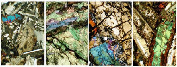
Light slows down as it moves into the mineral, and the two new polarizing vibration planes conduct light at different rates in the mineral. Each light ray also bends (refracts) as it passes in and out of the mineral, with an angle proportional to its change in velocity. Then they accelerate back to the same speed in the air out the other side. However, the light waves of the rays are now out-of-sync because they moved at different velocities within the mineral. When the wave planes are combined into one plane by the second filter, the waves "interfere" with each other, producing new wavelengths with startling new "interference colors." This amazing feature of birefringence (two refractions) is caused by the two mineral light rays. Birefringence and interference colors vary according to the particular crystal structure unique to each mineral, and so we can interpret important crystallographic properties and identify microscopic minerals, and often infer some of their chemical compositions.
Tools such as compensator wave plates can also provide clues to identification, while reference books provide data tables, optical descriptions, and methodology. There are many written for students and professionals, and you should have one or more. If the book is a few years or decades old, not a problem, as the technique has not changed much, and the minerals not at all! My own library is rather dated, actually. Phillips has several chapters on the use of the universal stage, while Kerr has a fine section on mineral characteristics. Ehlers is one of the most comprehensive resources, and petrography/atlas books are great for illustrating what you actually see. These favorites are:
Optical Mineralogy by Paul Francis Kerr (1977, McGraw Hill)
Optical Mineralogy by Ernest Ehlers (in two volumes, 1987, Blackwell Scientific)
Mineral Optics by Wm. Revell Phillips (1971, Freeman)
Petrography of Igneous and Metamorphic Rocks by A. R. Philpotts (2003, Prentiss Hall)
Atlas of Rock-Forming Minerals in Thin Section by Mackenzie and Guilford (1984, Longman)
Any crystalline material might reveal features of its structure and chemistry via PLM, including wonderful new colors. Not just inorganic rocks and minerals, but also hard parts of plants and animals, and organic crystals formed by diseases. Interesting and beautiful effects might appear as well as new details, so it is worth a look for almost any sample you are studying. And that is easy to do with a pol scope.
A few images from my basalt slides are below, showing interference colors typical of common birefringent minerals. See this Idaho State University page about thin sections of rocks, which since the late 1800s have provided the evidence for much of what we know about the Earth. Where would science be without microscopes?
Although all the major makers produce models of polarizing microscopes (see below), there has been some decline during recent years in their use in geological education and research. Considering that the Earth is made of rocks, and rocks cannot be studied or understood without petrographic microscopes, this is not good! See this article by Mickey Gunter about the problem.
Greg's Leitz Microscopes Page
Here is a separate page about Leitz Wetzlar microscopes, including photos, parts, accessories, documents, and links. All types, not just pol scopes. Other brands (including Leica) will stay on this page for now. These are files that I have collected or borrowed and saw a need to organize. There is not much before the large models of the 1960s and later, so please send me corrections and additions of documents to help the page grow. Model names, variations, and options for Leitz microscopes are complex and confusing -- it would be nice if someone (not me) started a Leitz microscopes Yahoo Group or website.
Greg's Olympus Microscopes Page
Here is a separate page about Olympus microscopes, especially the BH2 line, including photos, links, and documents. Biological too, not just pol types. I have a lot of documents and photos of my favorite models from Olympus. This page is mainly about the CH, CH2, BH, and BH2 models, plus some older ones, and a little bit about the BX models that were introduced in the 1990s.
You might also be interested in my new Facebook Page: Greg's Microscopes


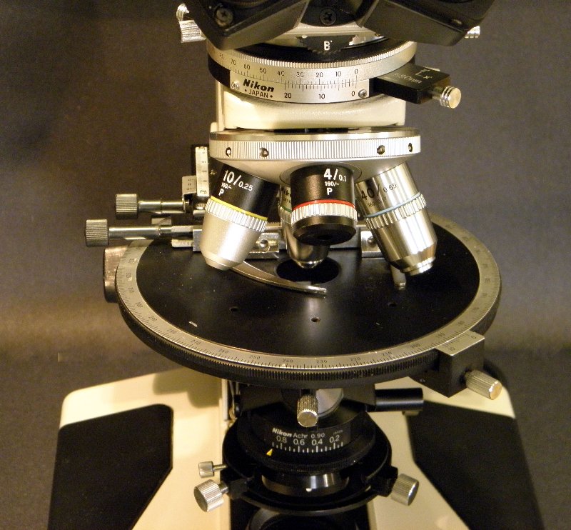
Several important features common to petrographic microscopes are illustrated in this photo of the middle section of my Olympus BH2 pol scope. Between the head and the arm is an intermediate tube with a Bertrand lens, an analyzer filter, and 6x20 mm slot for a compensating wave plate. The Bertrand lens has a dial to swing it in and out of the light path, with a second dial to focus it. The analyzer (upper polarizing filter) can be moved in and out via a sliding plate, and its part of the tube is marked in degrees for rotation, with a locking screw. Wave plates for the open slot have notches to allow precise stops for an open hole or the wave filter.
The nose turret slides on a dovetail for easy replacement, and holds four DPlan objectives (here 4x, 10x, 20x, 40x) designated PO for their strain-free construction. Three of the turret holes can be centered via side screws, using special small wrenches, or because these are easily lost and hard to replace, small screwdrivers. The same wrenches fit two centering screws for the circular stage, which is graduated in degrees with a lever to engage a "click" every 45 degrees, and a stop screw to lock the stage in place. A special x-y mechanical stage (slide holder) is made with low control knobs, so as to not interfere with the objectives during rotation. It is removable because some petrographers prefer to move slides by hand.

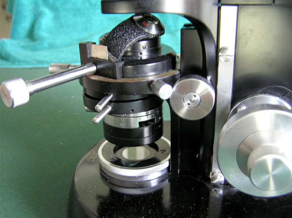
Beneath the stage is a Pol (strain-free) condenser with a flip-up top lens, n.a. 0.25/0.90. This top lens concentrates light for the higher-power objectives, needed when the Bertrand lens is used to make a "conoscopic" view of an "interference figure" (a diagnostic light pattern formed on the the back lens surface of the objective). The lower part of the condenser holds a polarizing filter that can be rotated, with a click for 90-degree "crossed polarizers." On my Olympus, all of these special pieces are very well made, and I take good care of them because they are expensive!
Glass can become slightly polarizing under uneven pressure or if it is not cooled evenly, leaving residual "strain." Because there is usually a pol filter in the light path (under the condenser), an extra source of polarization will add unwanted birefringent effects, so optical parts are constructed (or carefully selected from normal production) to minimize strain. Such objectives, eyepieces, and condensers might be marked P, Po, Pol or SF (strain free), or have red lettering. However, many "regular" optical parts might work OK in your pol scope as well, so it is worth a test. A bigger problem is reflection polarization by mirrors and prisms in binocular and trinocular heads, which is why many older pol scopes use monocular heads. Two-eye viewing is worth some extra cost if you can find a binocular pol head, or if not, you might be willing to put up with slight induced birefringence from a standard biological head. Of course, there will be none if you can swing out or remove both of the pol filters when viewing in "plain" light.
What do they cost?
Because they were made in relatively small numbers, or their modular parts could be rather specialized, petrographic microscopes both new and used command premium prices -- often from double to triple the price for a biological version of the same model, depending on how complete it is. A few BX pol models have been seen on eBay for $6000 to $12,000. BH2 BHTP models that are complete enough to use go for $2500 and up, but sometimes partial stands that need work appear on eBay for $1000-$1500, depending on what parts they have. Older models (1960s-70s) of other brands are usually less than $1500, with student models often well below $1000. These tend to be monocular, sometimes binocular, and trinocular heads may exist for several hundred $ more. But -- be sure these are pol versions. The monocular "horse shoe foot" models of the 1960s and earlier (and more recent Chinese versions) are often around $300 to $700. Look for examples with wave plates and at least 3 objectives.
If you are outfitting one with additional optical parts, you might find that less expensive objectives, eyepieces, and even condensers from biological (i.e. not certified as strain free) versions work just fine, or at least are so close that their strain effects are hardly noticeable. But if you are buying one at its higher petrographic price, it should already have the proper strain-free versions of optics to get your money's worth (and to preserve its higher resale value).
For several years I counted items with "microscope" in their titles as listed on eBay.com, by brand and type. See eBay Microscopy (an excel spreadsheet) for a table and charts of changes by month in these numbers. A high total near 10,000 items was reached in August 2009, but as the recession eased, the number of microscope listings dropped. Prices dropped too, but have since come back (most are still bargains, but watch out). In 2010, I stopped counting because eBay changed the way it lists items in my microscope categories, and the new counts can not be compared with my record. As you might guess, the big four are the most popular brands, but there are plenty of AO and B&L scopes and parts out there as well. eBay prices for microscopes remain at bargain levels, but there are now a lot more sold at fixed prices, whereas auctions of valuable items at low starting prices are no longer common.
Also see these notes about adapting digital cameras to my Olympus BH-2 and BX50 microscopes. See more about Olympus cameras on my Olympus BH2 web page. On eBay are my Guides to Buying Microscopes and Caring for Microscopes. If you find them to be useful, please check the response boxes so I can stay in the top 1000!
Below are galleries of polarizing microscopes both new and old, some interesting accessories, links to useful websites, and documents including pdf files of scanned instruction manuals. Please consider sharing your pdf documents and jpeg images related to polarizing microscopes (email to me). Thanks to these fine folks for permission to use their images and documents:
American Optical Spencer and Polarstar manual -- William Gasser
AO-Spencer documents on Steve Neeley's website
AO Pol Scopes Catalog -- Humboldt St. Univ. Scientific Instr. Museum
AO Spencer Pol #41 -- Jay Stanley of Classic Optics
B&L Dynoptic Pol scope -- Frank Nasser @ microlites.com
Cooke polarizing microscopes brochure -- Mark Glusker
Gillett & Sibert student scope -- Richard Morris (bluecerise400)
Science-Info website for microscope documents -- Gordon Couger
Swift and Son lab scope, and Wild M21 objectives -- Ray Sloss
Leitz Laborlux and SM Lux Pol scopes -- Ridge Equipment
Leitz Ortholux Pol scope and pol parts -- Tamagno
Leitz Ortholux camera and light meter -- William Day
Leitz Aristomet research scope -- Chip Sanders (mr-keyboard)
Leitz AM Pol and SM Pol scopes -- Raymond Hummelink
Leitz MOP image and 1913 catalog -- Charla Mason
Leitz HM Lux pol scope and brochures -- Allen Carpenter
Leitz older documents -- Don Grybeck
Lomo Min-8 benchtop scope -- Dirk Marel (eapoecistron)
Meopta and ROW images and manuals -- Werner Hartmann
Microscope documents from Gordon Couger's website
Nikon Ske pol scope -- Jay Stanley of Classic Optics
Nikon Alphaphot2 Pol -- John Woodhouse
Nikon Optiphot Pol manual -- Ian Hutcheon
Nikon Microphot Pol image -- petunia_d (eBay seller)
Olympus POM and Nikon POH-2 brochures -- Mike Symons
Olympus AH/BH/CH pol scopes brochure -- Jay Stanley of Classic Optics
Olympus Vanox -- Mark Home (microscopesolutions)
Reichert Diastar Pol -- Robert (zeissisnice1)
Reichert Zetopan -- electrowise
Unitron brochure -- Jason Weinstein
Zeiss Pol brochures and pricelist -- Paul V. Heinrich
Zeiss Ultraphot II scope -- Frank Nasser @ microlites.com
Zeiss and Leitz price list catalogs -- Jason Weinstein
Zeiss aus Jena benchtop scope -- BIGGERBY2002
About documents and images posted and linked below: Many are scans that I have made from my personal collection of old manuals and brochures, or are my photos of microscopes I have owned. Many others are posted by special permission but remain the property of the generous donors. I don't make money from this website -- it's a personal hobby. Images, documents, and web files might have no copyright notices, but that does NOT mean they are public property, as any corporate lawyer could tell you. Some unscrupulous vendors on eBay sell document scans without permission, but the ones here are for your personal reference only, not for sale or commercial use. If you or your company own rights to any of the files below, and you object to having them posted here, please let me know and I will remove them or their links immediately.
Documents, files, links, and images on these pages are for your personal use and information, and many are my property or the property of people who have allowed me to post them here. They are not for commercial use including eBay and may not be altered, sold, distributed, or otherwise used without specific permission from me or the people who own them.
Images of Polarizing Microscopes
Models on the Current Market (Fall 2012)
Hinotek polarizing microscopes
Models Produced in the Past
American Optical Spencer #41 research scope
American Optical Spencer P45 student scope (1960s)
American Optical (Reichert) Series 110 (Polstar)
Bausch&Lomb Dynoptic Polarizing scope model LI (1966)
Bausch&Lomb Dynoptic Polarizing scope model LM (c. 1966)
Cooke, Troughton, & Simms lab scope (1950s)
Gillett & Sibert student scope (1970s)
Leitz Wetzlar Microscopes (separate page link)
Lomo Min-1 packed in its lunch box
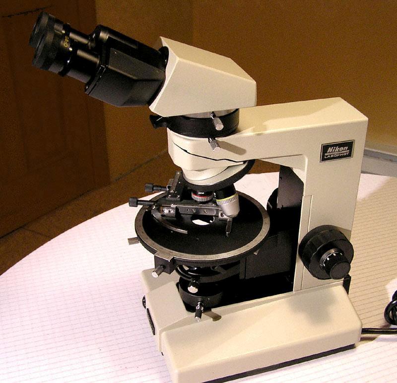
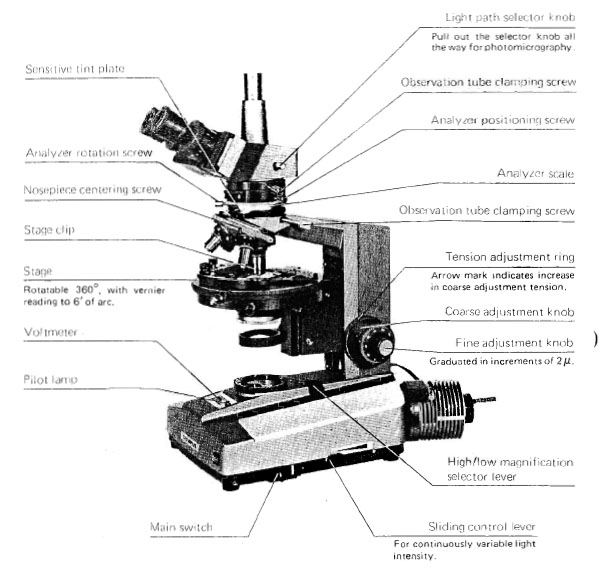
Nikon Labophot Pol
Nikon Microphot FX Pol (c. 1985)
Reichert Austria Neopan binocular
Reichert Austria small monocular
Reichert Austria benchtop scope
Reichert Zetopan research scope
Swift and Son (England) lab scope
Vickers M14/2 monocular lab scope
Vickers M15 monocular w/incident light accessory
Vickers M70 monocular w/incident light accessory
Wild Heerbrugg M5 Stereo Photo Viewer
Zeiss aus Jena Laboval 3 student scope
Zeiss aus Jena Amplival benchtop scope
Zeiss Standard 16 binocular (gray)
Zeiss Standard 14 monocular (gray)
Parts and Accessories
AO Spencer compensator plates and micrometer slide
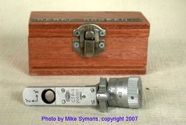
CTS incident light attachment w/objectives clip
Leitz Dialux Pol condenser c.1970
McCrone dispersion staining objective
Meiji MT9000 point-counting mechanical stage
Nikon Coolpix 990 with Leitz Periplan relay lens
Nikon pol objectives (as per Labophot)
Nikon Labophot/Optiphot middle section
Nikon Optiphot2 Pol intermediate tube
Nikon S Pol condenser and turret
Olympus POS objectives and accessories
Olympus POM objectives and accessories
Olympus POM Berek compensator (link to instruction manual)
Olympus BH "simple polarizing" filters
Olympus BHA-P circular stage with mechanical stage
Olympus BH2-SRG circular stage with C-FMP mechanical stage
Olympus BH2-SRP circular stage with A-FMP mechanical stage
Olympus BH2 Berek and wave compensator plates
Olympus BH2 Pol objectives and condenser
Olympus BH2-PA pol intermediate tube
Olympus BH2-KP simple pol intermediate tube
Olympus BHS-LSH 100 watt lamp house
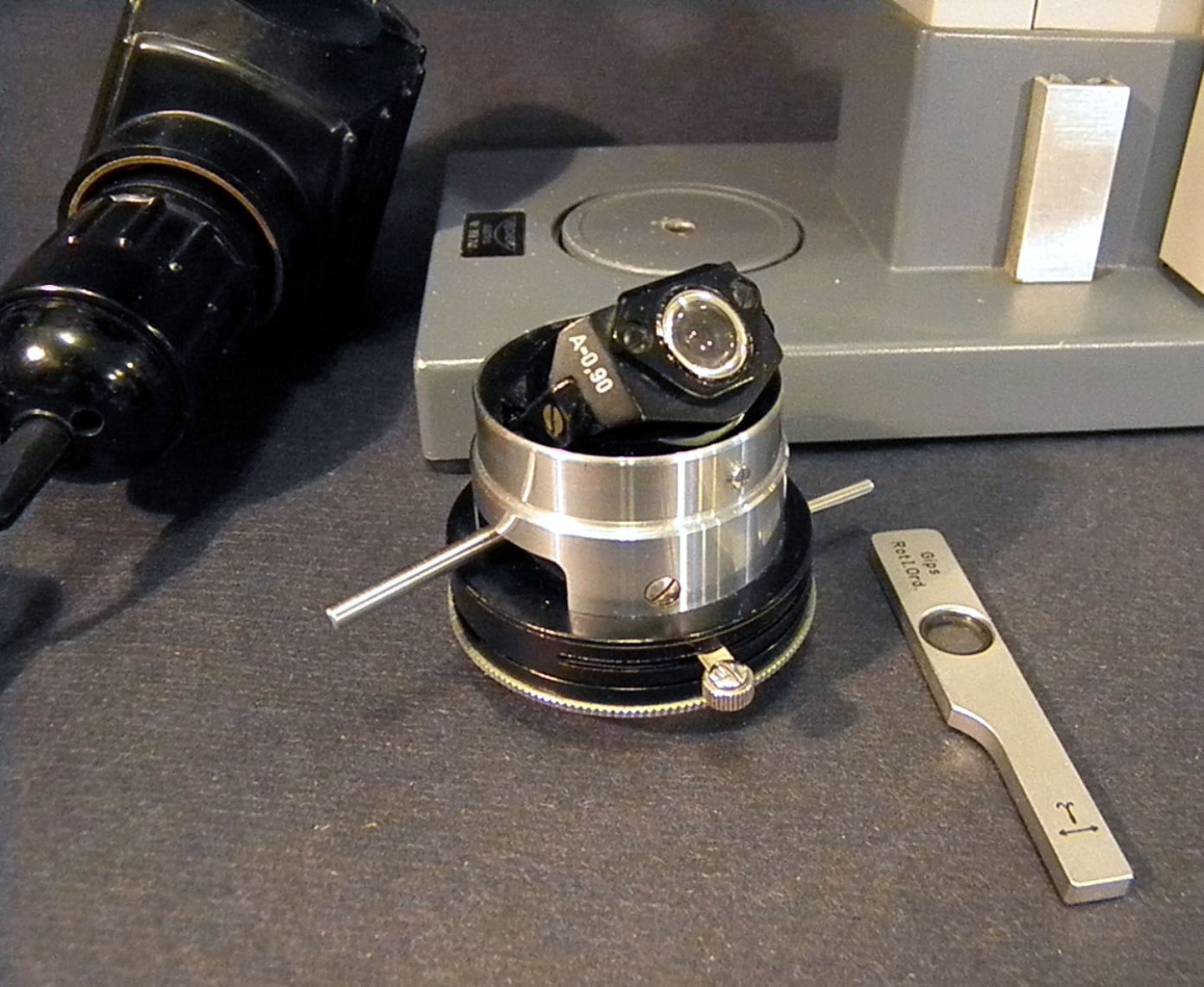
Reichert Neopan-Pol condenser, quartz plate, and lamp
Reichert Neopan-Pol objectives
Reichert Zetopan-Pol analyzer and compensator plates
Unitron MPS centering turret in clutch
Vickers compensating wave plates
Wild M21 pol objectives (centering barrels)
Wild M21 pol stage X-Y slide holder
Zeiss Jena intermediate tube and wave plate
Zeiss black monocular pol head
Zeiss Universal rotary stage w/centering wrench
Zeiss Universal Pol middle section
Zeiss pol objectives, 2-ring centering
Zeiss Standard analyzer plate in intermediate tube
Pol Scope Instruction Manuals and Documents
AO Polarstar Series 2300 reference manual
AO Spenser Polarizing Microscopes catalog, c.1950
AO Blue Book polarizing microscopes chapter, c.1950?
AO 110/120/Series 10 Microstar Analyzer/Polarizer Reference Manual
B&L Dynoptic Polarizing Microscope Instructions, 1966
B&L Polarizing Microscopes catalog, 1970
B&L Polarizing Microscopes price list, July 1970
Leica Polarization Use brochure
Leitz Dialux Pol catalog 55-16a, 1960s
Leitz 1960 Pol Scopes Catalog 55-20 with 1961 price list
Leitz 1971 Pol Scopes Catalog 550
Leitz SM-LUX Pol instructions 550-34
Leitz Orthoplan-Pol instructions (older gray version)
Leitz Orthoplan-Pol brochure (newer white version)
Leitz HM-POL instructions (1970s)
Leitz SM-POL Directions 55-19 (1960s)
Leitz Laborlux-Pol Directions 55-6 (1960s)
Leitz Laborlux Pol instructions (1960s model)
Leitz Laborlux 11 Pol instructions code 933 409 (1980s model)
Leitz Laborlux 12 Pol instructions code 933 407 (1980s model)
Leitz Laborlux 11 S Pol and 12 S Pol brochure (c.1990)
Leitz Dialux Pol Directions 55-12 (early 60s)
Leitz Dialux Pol Instructions 550-12a (late 60s)
Leitz Ortholux -Pol instructions 55-25a
Leitz Ortholux-2 Pol instructions
Leitz Panphot Polarizing Microscope with Photo Equip brochure
Leitz Waldman Hollow Glass Sphere instructions
Leitz Elliptical Compensator Directions
Leitz Berek Compensator Directions
Polarized Light Microscopy (Leitz, 3rd edition, 1985)
Meiji ML-POL instruction manual
Meiji ML-9000 series instruction manual
Meopta DRU instructions (1960s) in German
Nikon Eclipse E200 Pol Instructions
Nikon Labophot Pol Instructions
Nikon Labophot2 Pol Instructions
Nikon Optiphot Pol instructions
Nikon Optiphot Pol and Labophot Pol Brochure
Nikon Optiphot Pol and Labophot Pol Prices (June 1982)
Olympus POS instruction manual
Olympus POM instruction manual
Olympus BH-A-P instruction manual
Olympus AH/BH/CH Pol Scopes Brochure (transitional models)
Olympus BH-2 BHT-P instruction manual
Olympus BH-2 BHS-P instruction manual
Olympus CH CHA-P instruction manual
Olympus BHSP-BHTP-CHTP pol scope price list, 1992
Olympus Berek Compensator Instruction Manual (early 70s)
Olympus BX-Pol parts description (1994)
Olympus BX51P/41P instruction manual (2004)
Reichert Neopan-Pol operating instructions (1968)
ROW Poladun IV instructions (1960s) in German
Spencer 1943 'The Use of Polarizing Microscopes' on Neeley's website
Spencer Polarizing Microscopes catalog with 1948 price list
Unitron Bio-Pol instruction manual
Wild M21 Polarising Microscope Instructions for Use (larger version)
Wild M21 instruction manual (smaller version)
Zeiss Axioskop 40 Pol brochure
Zeiss Axiolab Pol operating manual
Zeiss polarizing microscopes price list c.1959
Zeiss Winkle polarizing microscopes brochure and parts, c.1959
Zeiss polarizing microscopes brochure c. 2005
Zeiss Standard Pol Operating Instructions
Zeiss Standard Junior Pol Operating Instructions
Zeiss Universal R Pol brochure
Zeiss Universal Instructions for Use
Zeiss Universal Parts Catalog (1978)
Zeiss Standard Intermediate Tube
Zeiss Standard Pol Accessories
Zeiss Polarizing Microscope Price List (1982)
Zeiss Polarizing Microscopes Catalog (1979)
Zeiss Student Polarizing Microscopes Catalog (1978)
Internet Articles and PLM Websites
Micscape article on "The Six Best Polarizing Microscopes"
Micscape article on "Student Petrographic Microscopes"
Microscopy UK's Micscape Magazine -- see the index for PLM articles
Gordon Couger's website for microscope documents
Molecular Expressions Optical Microscopy Primer
Nikon's MicroscopyU Introduction to Polarized Light
Olympus Polarized Light Microscopy Primer
Olympus Applications of Polarising Microscopy
Olympus Basics of Polarising Microscopy
Microscopy from the Very Beginning
Olympus Polarized Light Image Gallery
The Care and Cleaning of Optics
Zeiss "How to Clean Optical Components"
J. M Derochette's Microscopy and Minerals web site
DMOZ directory of microscope dealers and suppliers
Lightscapes Webring for PLM imagery
Guide to Thin Section Microscopy
P. S. Neeley's AO Spenser microscope page
Making fossil thin sections by hand (Richard Hill)
Making rock thin sections by hand (Greg McHone)
Making thin sections in a geology lab (Union College)
Logitech Thin Section Preparation
Hillquist petrographic machines and supplies
Zeiss Conoscopy and Michel-Lévy Interference Color Chart
Asbestos identification Methodology by PLM - NIOSH 9002
Asbestos Counting Methodology by PCM - NIOSH 7400
More about collecting and analyzing asbestos by PLM
Dispersion Staining article by Walter McCrone
Thin Section Preparation & Microscopy refs by rbridges
Geochemical Society issue with universal stage and pol puzzle


My previous Olympus BH2 BHT-P at its "station" and with a Nikon Coolpix 990 digital camera mounted for quick photography. Note the PM-10 automated 35 mm camera controller at right, which is connected to a film camera back that can be easily set in place of the digital camera. The table is a heavy mdf laminate top screwed onto a frame base made of 2x4 lumber, making a rigid and very solid work bench free of tremors.
Corporate Websites